AI Assisted "Virtual Biopsy": A New Era in Non-Invasive Prostate Cancer Diagnostics

Biopsies are a standard tool in urology, used to detect tumors (both benign and malignant), identify disease processes, and guide treatment decisions. They have become central to modern practice and are considered by international guidelines as the gold standard for diagnosis and prognosis in urological cancers.
However, conventional biopsies are invasive. They often take place separately from treatment, exposing patients to additional risks such as bleeding or infection. They may also provide limited or inconclusive results, since only a small portion of tissue is sampled.
Against this backdrop, the idea of a “virtual biopsy” has emerged. This refers to gathering detailed biological information from tissue without physically removing it. While sometimes dismissed as a trendy AI term, virtual biopsy represents a genuine shift: a future where diagnosis may move from invasive sampling to image-based and computational methods, with accuracy comparable to traditional biopsies.
Virtual biopsy offers several advantages:
- Non-invasive and repeatable: It uses imaging methods already available in clinical care, so no extra cost or risk to patients, and tests can be repeated multiple times.
- Whole-tumor assessment: Instead of examining just a small tissue sample, imaging allows the entire tumor to be studied. This is crucial in cancers like prostate, bladder, and kidney cancer, where different parts of the tumor can vary biologically.
- Broader accessibility: Imaging is usually possible for nearly all patients, whereas a traditional biopsy may be technically difficult or unsafe for some.
- Sustainability: Fewer repeat procedures and hospital visits make the process more efficient and reduce healthcare’s environmental impact.
Herein we break down what is needed to develop virtual biopsy technologies, focusing on urological cancers. We look at how tools like artificial intelligence can combine with i radiological and pathological imaging to improve lesion analysis, reduce invasive procedures, and ultimately improve outcomes for patients with urological cancers.

Developing AI-Based Virtual Biopsy Models
Modern AI—especially deep learning—has enabled automation of many diagnostic tasks. Earlier approaches relied on human-designed image features (handcrafted radiomics), but AI automatically extracts more optimal features from raw data.
.In practice, this involves::
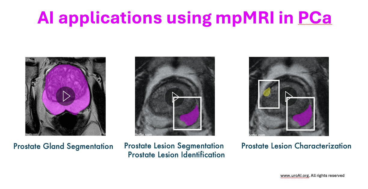
- Organ detection and segmentation: Identifying the target organ in an image to focus analysis and reduce noise from irrelevant regions.
- Lesion detection and segmentation: Highlighting abnormal areas within the organ, often combining multiple imaging methods.
- Simultaneous lesion segmentation and characterization Using AI to not only map the lesion but also grade its severity or significance.
To achieve this, AI models are trained on imaging data along with biopsy results, which serve as the “ground truth.” This allows AI to link imaging features to tumor grade, prognosis, or even genetic profiles. In this way, virtual biopsies can provide information once only possible through invasive sampling.
How Training with Ground Truth Works
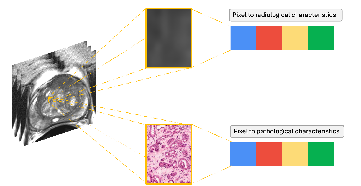
To train an AI model for virtual biopsy, we first need a reference standard, often called the ground truth. This ground truth usually comes from real biopsy results, where tissue samples are examined under a microscope and classified by experts (for example, cancer grade or tumor aggressiveness).
The AI model then studies medical images of those same cases (such as MRI or digital pathology slides) and learns to link the image features with the biopsy results. In other words, the biopsy provides the answer key, and the model learns the patterns in the images that correspond to those answers.
Once trained, the model can look at a new patient’s medical images and compare them to what it has learned from the ground truth cases. By doing so, it can estimate how likely the new lesion is to match certain diagnoses, such as tumor type, grade, or even genetic profile.
This process makes it possible for AI to “virtually biopsy” a lesion: drawing diagnostic conclusions from images alone, without the need to remove tissue.
How the Virtual Biopsy can make "the change"?
The Virtual biopsy may represents a major step forward in how we diagnose and manage urological cancers. By combining advanced imaging with AI, these methods make it possible to obtain diagnostic and prognostic information without physically removing tissue. This not only reduces the risks and discomfort associated with traditional biopsies, but also minimizes the number of unnecessary procedures.
At the same time, AI-driven analysis ensures that diagnostic performance can be pushed to the highest levels, offering accuracy comparable to—or even exceeding—that of conventional biopsies , possibly predicting more accurately the pathology from the radiology. Ultimately, these technologies expand access to care: they can be applied to a wider patient population, including those for whom biopsy is difficult or unsafe, and they streamline the diagnostic process by providing faster results.
The future is now: virtual biopsy has the potential to transform clinical workflows—reducing invasiveness, improving efficiency, and accelerating the path from suspicion to diagnosis—ultimately making cancer care both more effective and more accessible.

Some references that we found useful:
- Ronneberger, O., P. Fischer, and T. Brox. U-Net: Convolutional Networks for Biomedical Image Segmentation. in Medical Image Computing and Computer-Assisted Intervention – MICCAI 2015. 2015. Cham: Springer International Publishing.
- Chartier, S. and H. Arif-Tiwari, MR Virtual Biopsy of Solid Renal Masses: An Algorithmic Approach.Cancers (Basel), 2023. 15(10).
- Magoulianitis, V., et al., PCa-RadHop: A transparent and lightweight feed-forward method for clinically significant prostate cancer segmentation. Comput Med Imaging Graph, 2024. 116: p. 102408.
- Turkbey, B., et al., Fully automated prostate segmentation on MRI: comparison with manual segmentation methods and specimen volumes. AJR Am J Roentgenol, 2013. 201(5): p. W720-9.
- Mehralivand, S., et al., Multicenter Multireader Evaluation of an Artificial Intelligence-Based Attention Mapping System for the Detection of Prostate Cancer With Multiparametric MRI. AJR Am J Roentgenol, 2020. 215(4): p. 903-912.
